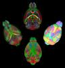
Johnson GA, Tian Y, Ashbrook DG, Cofer GP, Cook JJ, Gee JC, Hall A, Hornburg K, Kaczorowski CC, Qi Y, Yeh FC, Wang N, White LE, Williams RW. Merged magnetic resonance and light sheet microscopy of the whole mouse brain. Proc Natl Acad Sci U S A. 2023 Apr 25;120(17):e2218617120. doi: https://www.pnas.org/doi/10.1073/pnas.2218617120 Epub 2023 Apr 17. Erratum in: Proc Natl Acad Sci U S A. 2023 Jun 20;120(25):e2308718120. PMID: 37068254; PMCID: PMC10151475.
Merged magnetic resonance and light sheet microscopy of the whole mouse brain
Whole-Slide Cytometric Feature Mapping for Distinguishing Tumor Genomic Subtypes in Head and Neck Squamous Cell Carcinoma Whole-Slide Images
Blocker, S. J., Morrison, S., Everitt, J. I., Cook, J., Luo, S., Watts, T. L., & Mowery, Y. M. (2023). Whole-Slide Cytometric Feature Mapping for Distinguishing Tumor Genomic Subtypes in Head and Neck Squamous Cell Carcinoma Whole-Slide Images. Am J Pathol, 193(2), 182–190. https://doi.org/10.1016/j.ajpath.2022.11.004
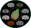
Prenatal heroin exposure alters brain morphology and connectivity in adolescent mice
Hornburg, K. J., Slosky, L. M., Cofer, G., Cook, J., Qi, Y., Porkka, F., Clark, N. B., Pires, A., Petrella, J. R., White, L. E., Wetsel, W. C., Barak, L., Caron, M. G., Johnson, G. A. (2023). Prenatal heroin exposure alters brain morphology and connectivity in adolescent mice. Nmr Biomed, 36(2), e4842. https://doi.org/10.1002/nbm.4842
Cytostatic hypothermia and its impact on glioblastoma and survival
Enam SF, Kilic CY, Huang J, Kang BJ, Chen R, Tribble CS, Ilich E, Betancur MI, Blocker SJ, Owen SJ, Buckley AF, Lyon JG, Bellamkonda RV. Cytostatic hypothermia and its impact on glioblastoma and survival. Sci Adv. 2022 Nov 25;8(47):eabq4882. https://doi.org/10.1126/sciadv.abq4882
Automated Nuclear Segmentation in Head and Neck Squamous Cell Carcinoma Pathology Reveals Relationships between Cytometric Features and ESTIMATE Stromal and Immune Scores
Blocker SJ, Cook J, Everitt JI, Austin WM, Watts TL, Mowery YM. Automated Nuclear Segmentation in Head and Neck Squamous Cell Carcinoma Pathology Reveals Relationships between Cytometric Features and ESTIMATE Stromal and Immune Scores. Am J Pathol. 2022 Sep;192(9):1305–1320. https://doi.org/10.1016/j.ajpath.2022.06.003
Late-onset cardiovascular dysfunction in adult mice resulting from galactic cosmic ray exposure
Bishawi M, Lee FH, Abraham DM, Glass C, Blocker SJ, Cox DJ, Brown ZD, Rockman HA, Mao L, Slaba TC, Dewhirst MW, Truskey GA, Bowles DE. Late-onset cardiovascular dysfunction in adult mice resulting from galactic cosmic ray exposure. Iscience. 2022 Apr 15;25(4):104086. https://doi.org/10.1016/j.isci.2022.104086
A time-course study of actively stained mouse brains: Diffusion tensor imaging parameters and connectomic stability over 1 year
Xiao, J., Hornburg, K. J., Cofer, G., Cook, J. J., Pratson, F., Qi, Y., & Johnson, G. A. (2022)
A time-course study of actively stained mouse brains: Diffusion tensor imaging parameters and connectomic stability over 1 year. Nmr Biomed, 35(1), e4611. https://doi.org/10.1002/nbm.4611
Resolution and b value dependent structural connectome in ex vivo mouse brain
Stephanie Crater, Surendra Maharjan, Yi Qi, Qi Zhao, Gary Cofer, James C. Cook, G. Allan Johnson, Nian Wang,
Resolution and b value dependent structural connectome in ex vivo mouse brain, NeuroImage, Volume 255, 2022, 119199, ISSN 1053-8119, https://doi.org/10.1016/j.neuroimage.2022.119199.
A multicontrast MR atlas of the Wistar rat brain
Johnson GA, Laoprasert R, Anderson RJ, Cofer G, Cook J, Pratson F, White LE. A multicontrast MR atlas of the Wistar rat brain. Neuroimage. 2021 Nov 15;242:118470. doi: 10.1016/j.neuroimage.2021.118470. Epub 2021 Aug 12. PMID: 34391877. https://www.sciencedirect.com/science/article/pii/S1053811921007436
Ex Vivo MR Histology and Cytometric Feature Mapping Connect Three-dimensional in Vivo MR Images to Two-dimensional Histopathologic Images of Murine Sarcomas
Blocker SJ, Cook J, Mowery YM, Everitt JI, Qi Y, Hornburg KJ, Cofer GP, Zapata F, Bassil AM, Badea CT, Kirsch DG, Johnson GA. Ex Vivo MR Histology and Cytometric Feature Mapping Connect Three-dimensional in Vivo MR Images to Two-dimensional Histopathologic Images of Murine Sarcomas. Radiol Imaging Cancer. 2021 May;3(3):e200103. doi: 10.1148/rycan.2021200103. PMID: 34018846; PMCID: PMC8183263. https://doi.org/10.1148/rycan.2021200103
A high-resolution interactive atlas of the human brainstem using magnetic resonance imaging
Conventional atlases of the human brainstem are limited by the inflexible, sparsely-sampled, two-dimensional nature of histology, or the low spatial resolution of conventional magnetic resonance imaging (MRI). Postmortem high-resolution MRI circumvents the challenges associated with both modalities. A single human brainstem specimen extending from the rostral diencephalon through the caudal medulla was prepared for imaging after the brain was removed from a 65-year-old male within 24 h of death. The specimen was formalin-fixed for two weeks, then rehydrated and placed in a custom-made MRI compatible tube and immersed in liquid fluorocarbon. MRI was performed in a 7-Tesla scanner with 120 unique diffusion directions. Acquisition time for anatomic and diffusion images were 14 h and 208 h, respectively. Segmentation was performed manually. Deterministic fiber tractography was done using strategically chosen regions of interest and avoidance, with manual editing using expert knowledge of human neuroanatomy. Anatomic and diffusion images were rendered with isotropic resolutions of 50 μm and 200 μm, respectively. Ninety different structures were segmented and labeled, and 11 different fiber bundles were rendered with tractography. The complete atlas is available online for interactive use [insert link to https://civmvoxport.vm.duke.edu/voxbase/login.php?return_url=%2Fvoxbase%2F]. This atlas presents multiple contrasting datasets and selected tract reconstruction with unprecedented resolution for MR imaging of the human brainstem. There are immediate applications in neuroanatomical education, with the potential to serve future applications for neuroanatomical research and enhanced neurosurgical planning through “safe” zones of entry into the human brainstem. https://doi.org/10.1016/j.neuroimage.2021.118135
Variability and heritability of mouse brain structure: Microscopic MRI atlases and connectomes for diverse strains
Wang N, Anderson RJ, Ashbrook DG, Gopalakrishnan V, Park Y, Priebe CE, Qi Y, Laoprasert R, Vogelstein JT, Williams RW, Johnson GA. Variability and heritability of mouse brain structure: Microscopic MRI atlases and connectomes for diverse strains. Neuroimage. 2020 Nov 15;222:117274. doi: 10.1016/j.neuroimage.2020.117274. Epub 2020 Aug 18. PMID: 32818613; PMCID: PMC8442986. https://www.sciencedirect.com/science/article/pii/S1053811920307606?via%3Dihub
Cytoarchitecture of the mouse brain by high resolution diffusion magnetic resonance imaging
Characterization complex collagen fiber architecture in knee joint using high-resolution diffusion imaging
Wang, Nian, Anthony J. Mirando, Gary Cofer, Yi Qi, Matthew J. Hilton, and G Allan Johnson. “Characterization complex collagen fiber architecture in knee joint using high-resolution diffusion imaging.” Magn Reson Med, January 21, 2020. https://doi.org/10.1002/mrm.28181.
Probing demyelination and remyelination of the cuprizone mouse model using multimodality MRI
Wang, Nian, Jie Zhuang, Hongjiang Wei, Russell Dibb, Yi Qi, and Chunlei Liu. “Probing demyelination and remyelination of the cuprizone mouse model using multimodality MRI.” J Magn Reson Imaging 50, no. 6 (December 2019): 1852–65. https://doi.org/10.1002/jmri.26758
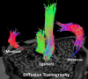
Whole mouse brain connectomics
Allan Johnson, G., Nian Wang, Robert J. Anderson, Min Chen, Gary P. Cofer, James C. Gee, Forrest Pratson, Nicholas Tustison, and Leonard E. White. “Whole mouse brain connectomics.” J Comp Neurol 527, no. 13 (September 1, 2019): 2146–57. https://doi.org/10.1002/cne.24560
Small Animal Multivariate Brain Analysis (SAMBA) - a High Throughput Pipeline with a Validation Framework
Anderson, R. J., Cook, J. J., Delpratt, N., Nouls, J. C., Gu, B., McNamara, J. O., Avants, B. Johnson, G. A., Badea, A. (2019). Small Animal Multivariate Brain Analysis (SAMBA) - a High Throughput Pipeline with a Validation Framework. Neuroinformatics, 17(3), 451–472. https://doi.org/10.1007/s12021-018-9410-0
Experimental influences in the accurate measurement of cartilage thickness in MRI
Wang, Nian, Farid Badar, and Yang Xia. “Experimental influences in the accurate measurement of cartilage thickness in MRI.” Cartilage 10, no. 3 (July 2019): 278–87. https://doi.org/10.1177/1947603517749917.
Neurite orientation dispersion and density imaging of mouse brain microstructure
Wang, Nian, Jieying Zhang, Gary Cofer, Yi Qi, Robert J. Anderson, Leonard E. White, and G. Allan Johnson. “Neurite orientation dispersion and density imaging of mouse brain microstructure.” Brain Struct Funct 224, no. 5 (June 2019): 1797–1813. https://doi.org/10.1007/s00429-019-01877-x
Quantitative susceptibility mapping as a biomarker for evaluating white matter alterations in Parkinson's disease
Guan, Xiaojun, Peiyu Huang, Qiaoling Zeng, Chunlei Liu, Hongjiang Wei, Min Xuan, Quanquan Gu, et al. “Quantitative susceptibility mapping as a biomarker for evaluating white matter alterations in Parkinson's disease.” Brain Imaging Behav 13, no. 1 (February 2019): 220–31. https://doi.org/10.1007/s11682-018-9842-z
Dynamic contrast-enhanced MRI promotes early detection of toxin-induced acute kidney injury
Privratsky, J. R., Wang, N., Qi, Y., Ren, J., Morris, B. T., Hunting, J. C., Johnson, G. A., Crowley, S. D. (2019). Dynamic contrast-enhanced MRI promotes early detection of toxin-induced acute kidney injury. Am J Physiol Renal Physiol, 316(2), F351–F359. https://doi.org/10.1152/ajprenal.00416.2018
Diffusion tractography of the rat knee at microscopic resolution
Diffusion tractography of the rat knee at microscopic resolution
Wang, N., Mirando, A. J., Cofer, G., Qi, Y., Hilton, M. J., & Johnson, G. A. (2019). Diffusion tractography of the rat knee at microscopic resolution. Magn Reson Med, 81(6), 3775–3786. https://doi.org/10.1002/mrm.27652
Accelerating quantitative susceptibility imaging acquisition using compressed sensing
Wang, N., Cofer, G., Anderson, R. J., Qi, Y., Liu, C., & Johnson, G. A. (2018). Accelerating quantitative susceptibility imaging acquisition using compressed sensing. Phys Med Biol, 63(24), 245002. https://doi.org/10.1088/1361-6560/aaf15d
Whole mouse brain structural connectomics using magnetic resonance histology
Wang, N., Anderson, R. J., Badea, A., Cofer, G., Dibb, R., Qi, Y., & Johnson, G. A. (2018). Whole mouse brain structural connectomics using magnetic resonance histology. Brain Struct Funct, 223(9), 4323–4335. https://doi.org/10.1007/s00429-018-1750-x
Imaging of Nanoparticle Distribution to Assess Treatments That Alter Delivery
Blocker SJ, Shields AF. Imaging of Nanoparticle Distribution to Assess Treatments That Alter Delivery. Mol Imaging Biol. 2018 Jun;20(3):340-351. doi: 10.1007/s11307-017-1142-2. Review. PubMed PMID: 29188418; PubMed Central PMCID: PMC6836466. https://link.springer.com/article/10.1007/s11307-017-1142-2
Compressed sensing in quantitative determination of GAG concentration in cartilage by microscopic MRI
Wang, Nian, Farid Badar, and Yang Xia. “Compressed sensing in quantitative determination of GAG concentration in cartilage by microscopic MRI.” Magn Reson Med 79, no. 6 (June 2018): 3163–71. https://doi.org/10.1002/mrm.26973
Diffusion tensor imaging using multiple coils for mouse brain connectomics
Nouls, J. C., Badea, A., Anderson, R. B. J., Cofer, G. P., & Allan Johnson, G. (2018). Diffusion tensor imaging using multiple coils for mouse brain connectomics. Nmr Biomed, 31(6), e3921. https://doi.org/10.1002/nbm.3921
Postmortem diffusion MRI of the entire human spinal cord at microscopic resolution
Calabrese, E., Adil, S. M., Cofer, G., Perone, C. S., Cohen-Adad, J., Lad, S. P., & Johnson, G. A. (2018). Postmortem diffusion MRI of the entire human spinal cord at microscopic resolution. Neuroimage Clin, 18, 963–971. https://doi.org/10.1016/j.nicl.2018.03.029
Susceptibility tensor imaging and tractography of collagen fibrils in the articular cartilage
Wei, Hongjiang, Eric Gibbs, Peida Zhao, Nian Wang, Gary P. Cofer, Yuyao Zhang, G Allan Johnson, and Chunlei Liu. “Susceptibility tensor imaging and tractography of collagen fibrils in the articular cartilage.” Magn Reson Med 78, no. 5 (November 2017): 1683–90. https://doi.org/10.1002/mrm.26882
Investigating magnetic susceptibility of human knee joint at 7 Tesla
Wei, Hongjiang, Russell Dibb, Kyle Decker, Nian Wang, Yuyao Zhang, Xiaopeng Zong, Weili Lin, Daniel B. Nissman, and Chunlei Liu. “Investigating magnetic susceptibility of human knee joint at 7 Tesla.” Magn Reson Med 78, no. 5 (November 2017): 1933–43. https://doi.org/10.1002/mrm.26596
Preferential adsorption of the additive is not a prerequisite for cononsolvency in water-rich mixtures
Wang, Jian, Nian Wang, Biaolan Liu, Jia Bai, Pei Gong, Geying Ru, and Jiwen Feng. “Preferential adsorption of the additive is not a prerequisite for cononsolvency in water-rich mixtures.” Physical Chemistry Chemical Physics 19, no. 44 (n.d.): 30097–106. https://doi.org/10.1039/c7cp04384h
An HPC Pipeline with Validation Framework for Small Animal Multivariate Brain Analysis (SAMBA)
Anderson, R. J., Cook, J. J., Delpratt, N. A., Nouls, J. C., Gu, B., McNamara, J. O., Avants, B. B., Johnson, G. A., Badea, A. (2017). An HPC Pipeline with Validation Framework for Small Animal Multivariate Brain Analysis (SAMBA). Corr, abs/1709.10483
Liposomal (64)Cu-PET Imaging of Anti-VEGF Drug Effects on Liposomal Delivery to Colon Cancer Xenografts
Blocker SJ, Douglas KA, Polin LA, Lee H, Hendriks BS, Lalo E, Chen W, Shields AF. Liposomal (64)Cu-PET Imaging of Anti-VEGF Drug Effects on Liposomal Delivery to Colon Cancer Xenografts. Theranostics. 2017 Sep 26;7(17):4229-4239. doi: 10.7150/thno.21688. eCollection 2017. PubMed PMID: 29158822; PubMed Central PMCID: PMC5695009. https://www.thno.org/v07p4229.htm
Influence of regional iron on the motor impairments of Parkinson's disease: A quantitative susceptibility mapping study
Guan, Xiaojun, Min Xuan, Quanquan Gu, Xiaojun Xu, Peiyu Huang, Nian Wang, Zhujing Shen, Jingjing Xu, Wei Luo, and Minming Zhang. “Influence of regional iron on the motor impairments of Parkinson's disease: A quantitative susceptibility mapping study.” J Magn Reson Imaging 45, no. 5 (May 2017): 1335–42. https://doi.org/10.1002/jmri.25434
Adult rat cortical thickness changes across age and following adolescent intermittent ethanol treatment
Vetreno, R. P., Yaxley, R., Paniagua, B., Johnson, G. A., & Crews, F. T. (2017). Adult rat cortical thickness changes across age and following adolescent intermittent ethanol treatment. Addict Biol, 22(3), 712–723. https://doi.org/10.1111/adb.12364
Exploring the origins of echo-time-dependent quantitative susceptibility mapping (QSM) measurements in healthy tissue and cerebral microbleeds
Cronin, Matthew J., Nian Wang, Kyle S. Decker, Hongjiang Wei, Wen-Zhen Zhu, and Chunlei Liu. “Exploring the origins of echo-time-dependent quantitative susceptibility mapping (QSM) measurements in healthy tissue and cerebral microbleeds.” Neuroimage 149 (April 1, 2017): 98–113. https://doi.org/10.1016/j.neuroimage.2017.01.053
Regionally progressive accumulation of iron in Parkinson's disease as measured by quantitative susceptibility mapping
Guan, Xiaojun, Min Xuan, Quanquan Gu, Peiyu Huang, Chunlei Liu, Nian Wang, Xiaojun Xu, Wei Luo, and Minming Zhang. “Regionally progressive accumulation of iron in Parkinson's disease as measured by quantitative susceptibility mapping.” Nmr Biomed 30, no. 4 (April 2017). https://doi.org/10.1002/nbm.3489
Magnetic susceptibility anisotropy outside the central nervous system
R Dibb, L Xie, H Wei, C Liu, Magnetic susceptibility anisotropy outside the central nervous system, NMR Biomed. 2017 Apr;30(4). PMCID: PMC5112155. https://onlinelibrary.wiley.com/doi/full/10.1002/nbm.3544
Joint 2D and 3D phase processing for quantitative susceptibility mapping: application to 2D echo-planar imaging
Wei, Hongjiang, Yuyao Zhang, Eric Gibbs, Nan-Kuei Chen, Nian Wang, and Chunlei Liu. “Joint 2D and 3D phase processing for quantitative susceptibility mapping: application to 2D echo-planar imaging.” Nmr Biomed 30, no. 4 (April 2017). https://doi.org/10.1002/nbm.3501

- Need help? 919-684-7755