Read the feature in Neuroscience News.com:… Read More

Small Animal Imaging Laboratory (SAIL)
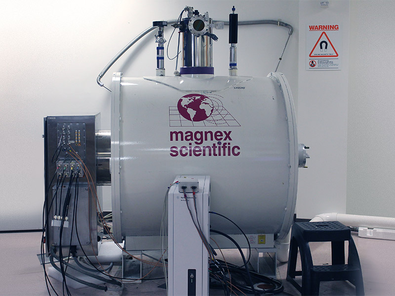
The CIVM, in collaboration with the Duke Pratt School of Engineering, a state-of-the-art Bruker 7.0 T preclinical MRI system. The system includes a range of radio frequency coils and a unique Resonance Research gradient coil providing more than twice the gradient strength of the majority of preclinical systems supporting higher spatial resolution and improved diffusion tensor imaging. Learn More
Duke Imaging Innovation Laboratory (DIIL)
With DIIL, CIVM is developing the next generation of imaging tools, leveraging the engineering expertise at Duke to push the technical boundaries of multimodal imaging. Featured equipment includes the 7 T Agilent MRI System and the 9.4T MRI System System. Learn More
Request a Project Collaboration
All project requests start here with your review of CIVM resources and rates and your submission of our online Project Request Form, either for SAIL or for DIIL. You'll be asked to provide the details of your project, including imaging needs, specific aims, timing and funding. Learn more and begin the project request form by choosing either SAIL or DIIL.
Recent News
The Duke Mouse Brain Atlas: New 3D Atlas Offers Unmatched Precision in Mapping the Mouse Brain
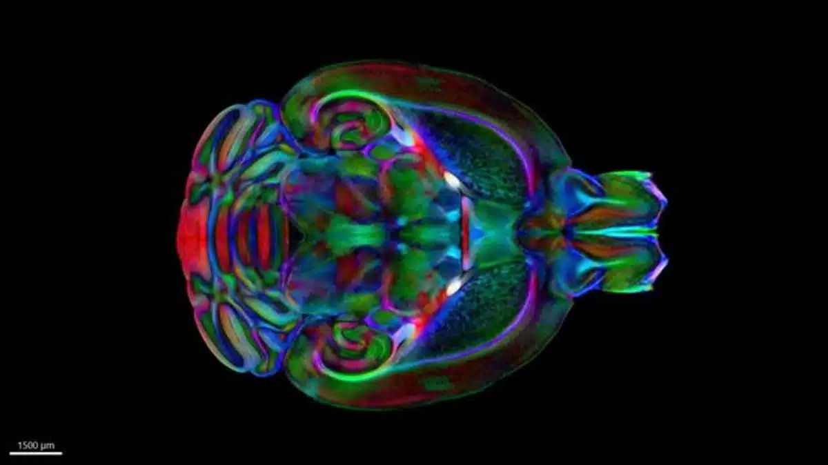
Brain Images Just Got 64 Million Times Sharper | Duke Today
DUKE TODAY - DURHAM, N.C. – Magnetic resonance imaging (MRI) is how we visualize soft, watery tissue that is hard… Read More
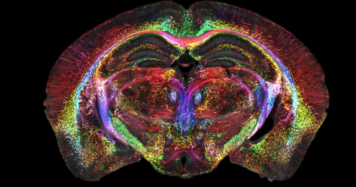
NeuroImage Vol No 255 chooses figure from Stephanie Crater's Paper for July Cover!
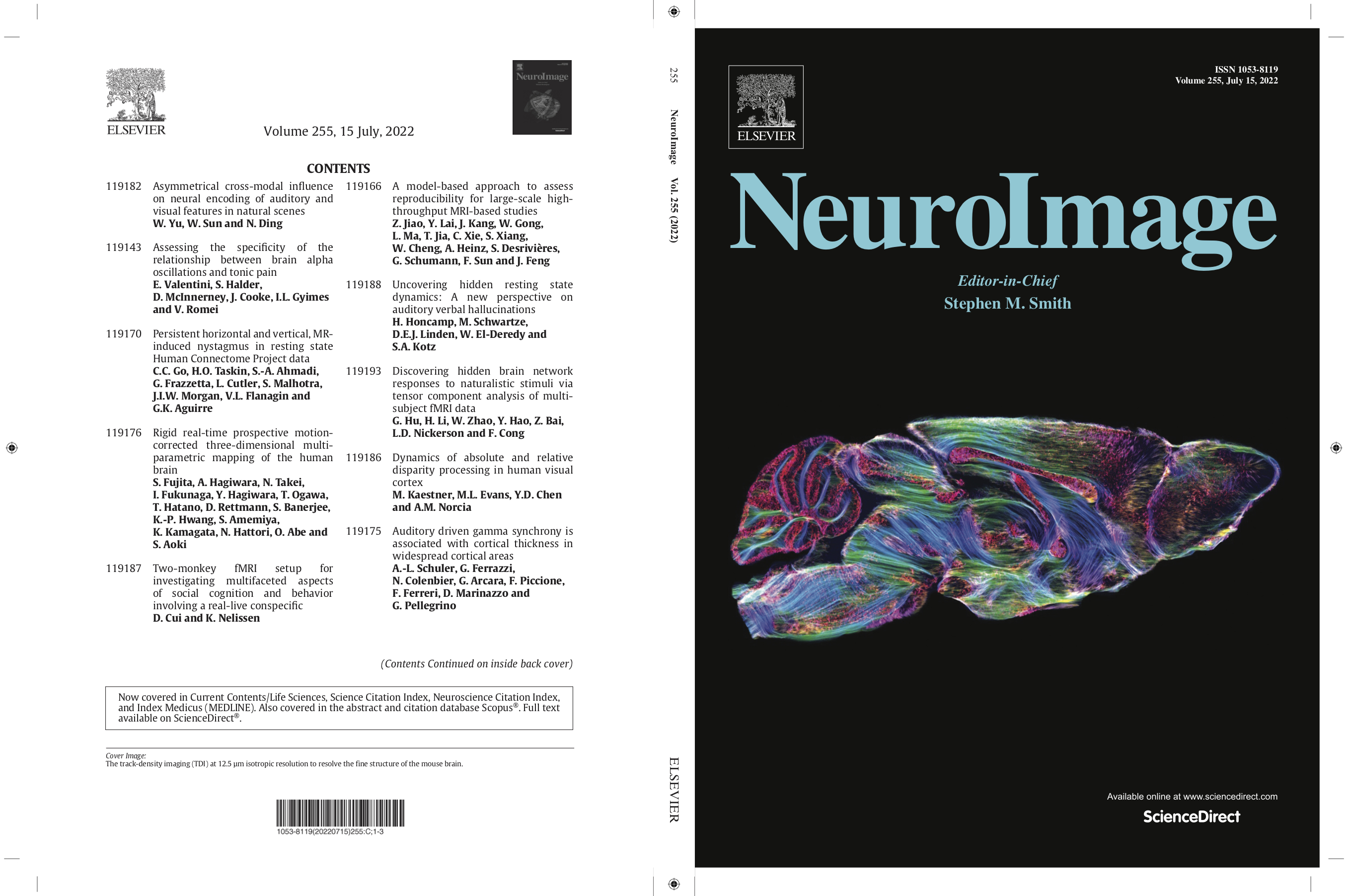
The cover in this issue of NeuroImage (Volume No 255) shows a representative image from a recent study by Wang et al… Read More
Recent Publications
The Duke Mouse Brain Atlas: MRI and light sheet microscopy stereotaxic atlas of the mouse brain.
Mansour H, Azrak R, Cook JJ, Hornburg KJ, Qi Y, Tian Y, Williams RW, Yeh FC, White LE, Johnson GA. The Duke Mouse Brain Atlas: MRI and light sheet microscopy stereotaxic atlas of the mouse brain. Sci Adv. 2025;11(18):eadq8089. Epub 20250430. doi: 10.1126/sciadv.adq8089. PubMed PMID: 40305623; PMCID: PMC12042906.
MR histology reveals tissue features beneath heterogeneous MRI signal in genetically engineered mouse models of sarcoma.
Blocker SJ, Mowery YM, Everitt JI, Cook J, Cofer GP, Qi Y, Bassil AM, Xu ES, Kirsch DG, Badea CT, Johnson GA. MR histology reveals tissue features beneath heterogeneous MRI signal in genetically engineered mouse models of sarcoma. Front Oncol. 2024;14:1287479. Epub 20240531. doi: 10.3389/fonc.2024.1287479. PubMed PMID: 38884083; PMCID: PMC11176416.
A rapid workflow for neuron counting in combined light sheet microscopy and magnetic resonance histology.
Tian Y, Johnson GA, Williams RW, White LE. A rapid workflow for neuron counting in combined light sheet microscopy and magnetic resonance histology. Front Neurosci. 2023;17:1223226. Epub 20230927. doi: 10.3389/fnins.2023.1223226. PubMed PMID: 37841684; PMCID: PMC10569694.
Merged magnetic resonance and light sheet microscopy of the whole mouse brain
Johnson GA, Tian Y, Ashbrook DG, Cofer GP, Cook JJ, Gee JC, Hall A, Hornburg K, Kaczorowski CC, Qi Y, Yeh FC, Wang N, White LE, Williams RW. Merged magnetic resonance and light sheet microscopy of the whole mouse brain. Proc Natl Acad Sci U S A. 2023 Apr 25;120(17):e2218617120. doi: https://www.pnas.org/doi/10.1073/pnas.2218617120 Epub 2023 Apr 17. Erratum in: Proc Natl Acad Sci U S A. 2023 Jun 20;120(25):e2308718120. PMID: 37068254; PMCID: PMC10151475.

- Need help? 919-684-7755