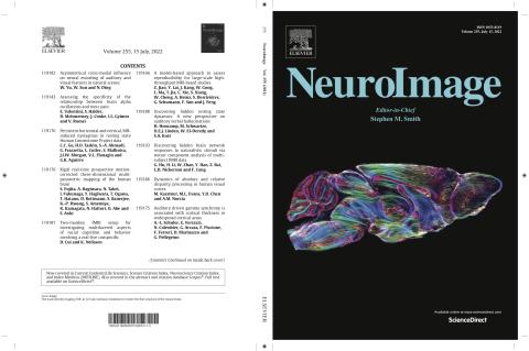
The cover in this issue of NeuroImage (Volume No 255) shows a representative image from a recent study by Wang et al from the CIVM that explores the tradeoffs in spatial and angular resolution and b values. The study has enables scientists at CIVM to obtain exceptionally quantitative connectomes at microscopic resolution.
Stephanie Crater, Surendra Maharjan, Yi Qi, Qi Zhao, Gary Cofer, James C. Cook, G. Allan Johnson, Nian Wang, Resolution and b value dependent structural connectome in ex vivo mouse brain, NeuroImage, Volume 255, 2022, 119199, ISSN 1053-8119, https://doi.org/10.1016/j.neuroimage.2022.119199.
Abstract: Diffusion magnetic resonance imaging has been widely used in both clinical and preclinical studies to characterize tissue microstructure and structural connectivity. The diffusion MRI protocol for the Human Connectome Project (HCP) has been developed and optimized to obtain high-quality, high-resolution diffusion MRI (dMRI) datasets. However, such efforts have not been fully explored in preclinical studies, especially for rodents. In this study, high quality dMRI datasets of mouse brains were acquired at 9.4T system from two vendors. In particular, we acquired a high-spatial resolution dMRI dataset (25 μm isotropic with 126 diffusion encoding directions), which we believe to be the highest spatial resolution yet obtained; and a high-angular resolution dMRI dataset (50 μm isotropic with 384 diffusion encoding directions), which we believe to be the highest angular resolution compared to the dMRI datasets at the microscopic resolution. We systematically investigated the effects of three important parameters that affect the final outcome of the connectome: b value (1000s/mm2 to 8000 s/mm2), angular resolution (10 to 126), and spatial resolution (25 µm to 200 µm). The stability of tractography and connectome increase with the angular resolution, where more than 50 angles is necessary to achieve consistent results. The connectome and quantitative parameters derived from graph theory exhibit a linear relationship to the b value (R2 > 0.99); a single-shell acquisition with b value of 3000 s/mm2 shows comparable results to the multi-shell high angular resolution dataset. The dice coefficient decreases and both false positive rate and false negative rate gradually increase with coarser spatial resolution. Our study provides guidelines and foundations for exploration of tradeoffs among acquisition parameters for the structural connectome in ex vivo mouse brain.
Keywords: HARDI; Diffusion MRI; Connectome; Crossing fiber; High-resolution